Dive into this comprehensive guide to explore kidney anatomy, physiology, and how to keep these incredible organs healthy. The kidneys are vital organs that perform life-sustaining functions, such as filtering blood, maintaining fluid balance, and excreting waste.
1) Normal Anatony:
Abdominal quadrant and it’s content abdominal organs by region adults kidney are like bee shape structure organ that are retroperitoneal is location they are normally located high in abdominal cavity and against its back wall lying on either side of the 12th Thoracic and third lumbar vertebral and outside the peritoneum the membrane that lines the abdomen and abdominal organs.
The kidney in the vetropertoneal cavity near the posterior body wall just below the diaphragm the lower ribs protect both kidneys the left kidney is located higher than the right kidney because of the size of the liver which occupy most of the right upper side quadrant of the abdomen.
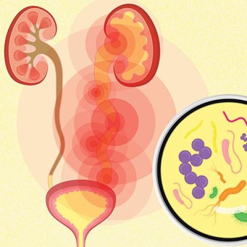
The urinary system consists of the right upper urinary tract kidney and ureter and the lower urinary tract bladder and urethra the functional unit of the kidney is the nephrons Relationship of the kidney supernal gland adrenaline and vascular structure to on another.
2) Physiology:
The kidneys are vital organs responsible for maintaining homeostasis. They perform several crucial functions:
- Filtration and Excretion: Kidneys filter blood, removing waste products and excess fluids. They excrete these substances in the form of urine.
- Reabsorption: Kidneys reabsorb essential nutrients, such as amino acids, glucose, and water, returning them to the bloodstream.
- Electrolyte Balance: Kidneys regulate the levels of ions, including sodium, potassium, and calcium, in the blood.
- Acid-Base Balance: Kidneys help maintain the body’s pH by excreting excess acid or base.
- Blood Pressure Regulation: Kidneys produce hormones that regulate blood pressure.
- Red Blood Cell Production: Kidneys stimulate the production of red blood cells by releasing erythropoietin.
Kidney Anatomy:
Anatomically, the kidney is enclosed by three layers:
- Renal Fascia: The outermost layer, providing structural support.
- Perirenal Fat Capsule: The middle layer, acting as a protective cushion.
- Renal Capsule: The innermost layer, directly surrounding the kidney and protecting it from infection.
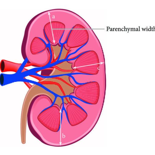
The renal medulla, the innermost part of the kidney, is responsible for urine concentration.
Renal Parenchyma:
Each kidney is composed of two main regions:
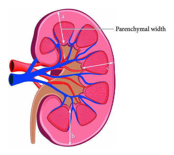
- Renal Sinus: The central cavity containing the renal pelvis, calyces, and renal blood vessels.
- Renal Parenchyma: The functional tissue of the kidney, divided into two parts:
- Renal Cortex: The outer region responsible for filtration of blood.
- Renal Medulla: The inner region responsible for reabsorption of water and solutes.
The renal pyramids, composed of renal medulla, are separated by renal columns, extensions of the cortex. The renal pelvis, a funnel-shaped structure, collects urine from the renal calyces. Urine flows from the renal papillae through the minor calyces, major calyces, and renal pelvis into the ureter.
Normal cortical thickness in adults ranges from 0.8 to 2.5 cm.
Location:
The kidneys are retroperitoneal organs located in the lumbar region of the abdomen. They extend vertically from the 12th thoracic vertebra to the 3rd lumbar vertebra. The right kidney typically sits slightly lower than the left due to the position of the liver.
Both kidneys are positioned posterior to the parietal peritoneum and anterior to the posterior abdominal wall muscles.
Shape size weight and orientation:
Each kidney is about 11cm broad and 3cm thick the left kidney is a little bit longer and narrower then the right kidney on an average the kidney weight is 150g in males and 135g in females the kidney are reddish brown in color.
Ureter:
The ureter are a pair of narrow ones thick wall muscular tubes which convey urine from the kidney to the urinary bladder they lied dead to peritoneum closely applied to the posterior abdominal wall in the upper part.
There are two locations of each kidney the size is 10 – 12 inches 30 – 25 cm long and the diameter ¼ inches 6_8 mm the location is behind the abdominal cavity
Anatomy and Physiology Function:
The ureter function is a transparent lots of time urine from the bladder to the kidney persistent urine back flow the kidney. The main part of ureter are is a Renal pelvis the collection of urine and proximal URETER behind the kidney.
Condition Of Ureteral:
- Stone is Ureteral
- Cancer in Ureteral
- Structure Ureteral
- Obstruction Uretro pelvis junction
Issues:
The main issues of ureter disturbance is:
- Severe pain
- Fever
- Fatigue
- Vomiting
- Nausea
- Blood In Urine
- Abdominal Pain
- Spinal Pain
The treatment options is surgery endoscopy and mediation.. the doctor for patients is intake lots of water because of reasons healthy lifestyle or healthy kidney
Physiology function:
- Excretory function
- Regulatory functions
- Synthetic function
- Endocrine function
Excretory function:
- Excretory wastes products especially nitrogenous and S-containing end products of protein metabolism
- Eliminate drugs and toxic substances and from the kidney function body
Regulatory functions:
- Help’s to maintain normal H ion pH and acid base
- Helps to maintain Na ion and electrolytes balance of body
- Helps to maintain osmotic pressure on the body and blood vessels and tissues fluids
- Helps to maintain water balance plasma volume and ECF extra cellular fluid volume
- Help to maintain blood pressure
Synthetic function:
Kidney synesthesia new substances like:
- Ammonia
- Hips uric acids
- Inorganic phosphate
- Gluconeogenesis
- Anatomy and Physiology
Endocrine function:
JG cell secrets Rennie in response to decrease renal arterial pressure and increase renal nerve discharge B1 effect
- Convert 25 hydrocolecaliferol to 1a 25 dihydrocolecalciferol ( vitamin D3 produce to kidney )
- Secretes erythropoietin in response to hypoxia RBC ( Red blood cells)
- Secret to prostaglandin that vasodilate the afferent arterioles to increase GFR glomerular filtration rate
Sonological Pathology of Kidney:
- Nephritis
- Stones in kidney
- Mass in kidney
- Hydronephrosis
Nephritis:
The inflammation of kidney is called nephrolithiasis
Cause:
- Bacterial infections E.coil
- Irritation (This is usually of stone)
- Cystanic infection ( sometime the infection of the kidney maybe cause by the cystitis mass
Clinical features:
Sever pain and tenderness in the lumber region
- High grade fever
- Nausea and vomiting
- Burring micturition
- Discharge of urine from urinary bladder
- Discharge of piss cell & protein In urine
Sonological finding:
In ultrasound we find inflammation in grade I parenchyma changes and grade II & grade III parenchyma changed
Masses In Kidney:
We maybe findthe following types of masses in kidney
- Solid masses
- Cystitis mass
- Cystic mass with internal echoes
- Complex mass teratomas
Solid Masses:
Often we find solid mass in the kidney but this mass might be possible single or multiple and if different size. The outer margin of the mass are mostly which is suggestive for benign tumor or nephroma but sometimes irregular masses seen which is suggestive for malignant tumor or Nephron carcinoma
Kidney Stones:
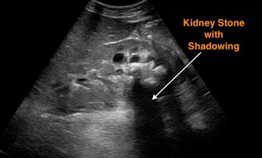
The pressure of stones in kidney is very common called nephrolithiasis it’s mostly in seen in cortical region the stones maybe single or multiple or in different sizes. The stone may be present in upper middle lower portion of the kidney in majority of the patients
Clinical Features:
- High grade fever
- Abdominal pain
- Back muscles pain
- Vomit
- Hematuria blood in urine
- Stuck problems in urine


